Pathogenesis of Canine Hip
Dysplasia
A
recently invented diagnostic method for canine hip dysplasia (1) was tested in
controlled studies and found to be accurate at 8-months of age. The dorsolateral
subluxation of femoral heads (DLS) test was devised to evaluate displacement of
femoral heads from acetabula when a hip is radiographed in a load-bearing
position (Fig. 1).
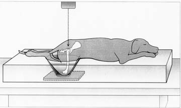
Figure 1: Illustration of a tranquilized dog in the sternal-abdominal position
on a foam rubber matt for the DLS test. The stifles and the ankles are bound
with tape. Care is taken to check for symmetry as viewed from the top and side.
Careful initial positioning is needed to avoid superimposition of the stifles
over the hip joints on the radiographic projection. The pad is about 5 inches
high for many dogs; a 4 inch or 3 inch thick pad may be used for smaller dogs to
ensure weight bearing on the stifles. The dotted line with the arrow head
represents the direction of the X-ray beam during the dorsoventral radiographic
procedure
The
test is done on anesthetized or tranquilized dogs positioned on their abdomen in
a kneeling position in a foam rubber pad so that body weight is transferred to
the bent stifles on the tabletop. A DLS score is calculated as the percent of
the femoral head covered by the acetabulum on a dorsoventral radiograph of hips
(Fig. 2).
|
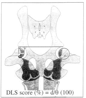 |
Figure 2: Illustration showing
how the DLS score is calculated from a dorsoventral radiograph. A straight
horizontal line is drawn between the acetabular lateral margins. A
perpendicular line is dropped from this line at the inside edge of the
femoral head and from the lateral edge of the acetabulum. The distance (d)
between these two perpendicular lines is measured in millimeters. The DLS
score expressed as percent is determined by dividing "d" by the diameter
of the femoral head (q, in mm) and multiplying by 100. The DLS score
represents the percent of the femoral head covered by the acetabular rim. |
In two
published reports (1, 2) dysplastic joints had a DLS score of 40% or less,
whereas normal hip joints had scores greater than 60% (see Figs. 3 and 4).
|
 |
 |
|
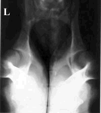
Figure 3: A
photograph of a dorsoventral radiograph of the position used for the DLS
test. The DLS score is determined on clear plastic overlays with the
radiograph. This dog had a DLS score of 40% for the left hip and 34% for
the right hip. "L" marks the left side of this dysplastic dog as viewed
from above the dog |
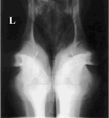
Figure 4: A
photograph of a dorsoventral radiograph of the weight bearing position
used for the DLS test. The DLS score is determined on a clear plastic
overlay with the radiograph. This dog had a DLS score of 63% for the left
hip and 66% for the right hip. "L" designates the left side of the
disease-free dog as viewed from above the dog. |
We
examined dogs at 8-months of age and related the DLS score to the appearance of
osteoarthritis (OA) at a later age (3). The presence of hip OA is an outcome
assessment of the dysplastic trait. Eight months was the preferred age since the
DLS score was stable by age 8-months, and 8-months might be early enough for
decision making for many breeding programs. Data suggested that the likelihood
ratio for developing an osteoarthritic hip joint for a DLS score > 55% = 0.2,
DLS score 45-55% = 2.6, and DLS score <45% = 8.0. Thus the likelihood ratios for
the presence of OA for the three DLS categories varied by a factor of 40 (3). We
have proposed that a dog with a DLS score of less than 55% at 8 months of age
has the dysplastic trait, In a follow-up study it was substantiated that dogs
with DLS > 55% at 8-months had normal hip joints on radiographs at two years of
age; 67% of dogs with DLS <50% at 8-months had hip OA on radiographs at two
years of age.
We
compared the DLS score and the PennHipTM distraction index (DI) at 8-months of
age to predict hip OA (4). Less than 40% of the variation in DLS score was
accounted for by DI; r2 = 0.36. A logistic multiple regression analysis was done
on the data and suggested that the DLS score had a better odds ratio for OA
joints to normal joints by a factor greater than two. The DLS score was more
accurate (had better sensitivity) than the DI for identifying hip OA, since the
DLS score gave low percentages of false-negative tests in the abnormal dogs.
Accuracies for predicting disease-free hip joints were similar for the two
methods (similar specificities) since both DLS score and DI gave low percentages
of false-positive tests in normal dogs (4).
Taken
together our results led us to conclude that the DLS score at 8-months of age
was a good predictor of hip osteoarthritis. Since both sensitivity and
specificity were high, the DLS score might have broad application for
identification of both disease-free and osteoarthritic hips. The DLS score is
useful at 8 months of age for programs selecting for either hip dysplastic or
normal dogs.
Literature References
1) Farese, J.P., Todhunter, R.J., Lust, G., Williams, A.J., and Dykes, N.L.
1998. Dorsolateral subluxation of hip joints in dogs measured in a
weight-bearing position with radiography and computed tomography. Vet. Surg.
27:393-405.
2) Farese, J.P., Lust, G., Williams, A.J., Dykes, N.L., Todhunter, R.J. 1999.
Comparison of measurements of dorsolateral subluxation of the femoral head and
maximal passive laxity for evaluation of the coxofemoral joint in dogs. Am. J.
Vet. Res. 60:1571-1576.
3) Lust, G., Todhunter, R.J., Erb, H.N., Dykes, N.L., Williams, A.J.,
Burton-Wurster, N., and Farese, J.P. 2001. Repeatability of dorsolateral
subluxation scores in dogs and correlation with macroscopic appearance of hip
osteoarthritis. Am. J. Vet. Res. 62:1711-1715.
4) Lust, G., Todhunter, R.J., Erb, H.N., Dykes, N.L., Williams, A.J.,
Burton-Wurster, N., and Farese, J.P. 2001. Comparison of three radiographic
methods for diagnosis of hip dysplasia in 8-month-old dogs. Jour. Am. Vet. Med.
Assoc.
219:1242-1246.