canine hip DySPLASIA
SUMMARY
Dysplasia coxofemoral is the bad formation of the articulations coxofemorais,
happening in all of the races, mainly in the big ones and of fast growth.
His/her transmission is hereditary, recessiva,intermitente and poligênica.
Nutritional factors, biomecânicos and of environment, associates to the
hereditariness, worsen the condition of the dysplasia. The suspicion to the
clinical exam is possible, but it is the study radiográfico, usually starting
from the twelve complete months of age in most of the races, by correct
positioning of the animal, that it defines so much diagnóstico.Para the
patient should be free from any reação.Este is reached with the general
anesthesia, preferably. The patient should be positioned in number decubitus,
members extended subsequent caudalmente, of equal length, parallel amongst
themselves and in relationship á spine, rotacionados medialmente, in such a
way that the patelas are put upon to the furrows trocleares. The pelve cannot
be tilted. In the minimum identification of the film it should consist the
number of registration of the dog, date of birth and it dates from the exam
radiográfico. the subluxação, usually as first sign radiográfico, can take to
the secondary artrose, denominated like this if it develops secondarily to
another alteration, in the in case displasia.O controls of this bad formation
is done through a selection radiográfica of all the animals used in the
reprodução.O index of Norbeg is used for diagnóstico.Modernamente the
treatment medicamentoso has if based on products with capacity anabolizante of
the cartilage to articulate degenerate.
A
diagnosis subject?
An appropriate clinical exam is not enough for the diagnosis of the dysplasia.
Definitively it will be radiográfico, by quality image and animal correctly
positioned.
Concept:
it is the bad formation of the articulations coxofemorais. Index in all of the
races, mainly in the big ones and of fast growth. He/she reaches the two
sexes, could commit an articulation (approximately 10%) or both.
Histoy:
Schnelle (1936) it described the dysplasia coxofemoral and Konde for the first
time (1947) it commented on his/her hereditary origin. Schales (1959) it
described her as bad formation and it indicated the exam radiográfico for the
diagnosis. Wayne and Riser(1964) they related the fast and precocious growth
and earnings of weight of German shepherds with genetic transmission.
Henricson,Norberg and Olsson (1966) they considered her as bad hereditary
formation and the subluxação as a consequence of the anatomical alteration.
Transmissão:
hereditária,recessiva,intermitente and poligênica (some authors have been
considering 20 genes). nutritional Factors, biomecânicos and of environment
(multifatorial), associates to the hereditariness, worsen the condition of the
displasia.Recomenda-if fundamentally to avoid the traumas, be them of the
obesity, of the places escorregadios,etc...
Etiopatogenia:
as
structures that aid in the maintenance of the articulations are: capsule to
articulate, ligament transverse acetabular, musculature of the area, round
ligament, negative pressure to articulate and application of the acetábulo for
the lip glenoidal or ligament acetabular.Pesquisadores has been basing their
studies in the biochemical modifications of the liquid sinovial, as the
decrease of the negative cloro(carga) and increase of the sodium and potassium
(positive loads). In function of these alterations an increase of the
osmolaridade, that brings as consequence the increase of the amount of the
same liquid and the sinovite with dehydration of the cartilage to articulate
happens. Starting from this instant a sequence of other episodes is uncoiled,
such as: I increase of the pressure intra to articulate, I increase of the
tension on the soft structures that it maintains the articulation, loosening
of these soft fabrics, loss of the intimacy to articulate, arrasamento
(ocificação or calcificação)ou not of the cavity acetabular (medial aspesto),
subluxação (lateral displacement of the femoral head, usually as first sign
radiográfico), edema, rupture partial or total of the round ligament, personal
computer fractures criminal acetabulares and finally the secondary artrose
(secondary because he/she grows secondarily to another alteration - the
diplasia). There is to still consider the hypothesis that the dysplasia is a
bad formation biomechanics, resulting from a disparity between the development
of the pelvic muscular mass and the fast growth of the esqueleto
(Illustration 1).
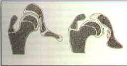
Illustration 1.
To the left drawing of an articulation normal and
anomalous
coxofemural to the right. Forces articulate abnormal they tilt the head and
femoral lap down,
they modify the anatomy of the cavity acetabular,rompem the round ligament and
they thicken the femoral lap.
Symptomatology:
it happens mainly among the four months until less than a year of life. The
dogs can present dificul-dades to get up, to walk, to run, to jump and to go
up stairways. The locomotion can be hindered in flat places. To run they can
imitate the race of rabbits. The claudicação can affect an or two members. In
the second case it is observed, with some frequency, that the animals move the
more weight on the previous members, developing the musculature thoracic
desproporcionalmente in relation to the subsequent ones. The last ones can be
shorter, could happen reluctance to the exercises, being observed preference
by the to sit down or to lie down. Abnormal episodes of aggressiveness are
sometimes observed, besides with the proprietor. The dysplasia can provoke a
lot of pains, to walk imperfeito,afetando the resistance of the animal.
Exam
clínico:
it´s made by
the observation of the animal in station, walking and trotting, in the
verification of increases of volumes and asymmetries and in the search of the
presence of the pain, crackling and width of the movement to articulate,
larger in the sharp and smaller phase in the chronicle, already in this last
one they intensify the alterations articulate degenerative, taking place the
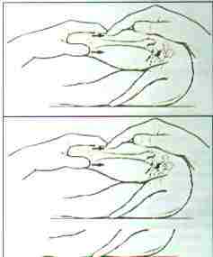
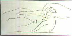
capsular and muscular fibrosis surrounding. The signs Ortolani and Bardens
should be explored in young dogs, anesthetized and put in lateral decubitus.
For for the sign of Ortolani (Illustration 2), position the femur superior
perpendicularmente to the longitudinal axis of the pelve and parallel to the
surface of the exam table. Put the palm of one of the hands on the
articulation coxofemoral under evaluation and with the another it holds the
articulation corresponding fêmoro-tíbio-patelar firmly, pressing the femur
against his/her acetábulo. When this pressure is exercised, the femoral head
of the articulation dysplastic subluxa back-sidelong. Maintain this pressure
and abduct to the maximum the femur. During this maneuver you will feel the
head of the femur will return his/her cavity acetabular, sometimes emitting an
audible sound similar to a" clunk." The return with or without sound it is
found clinical that corresponds to a sign positive Ortolani, coming to confirm
the presence of laxity to articulate. For the sign of Bardens (Illustration
3), suitable for lighter animals and with less than three months of age, hold
the superior femur with a hand and position the other with the thumb in the
tuberosidade isquiática, the indicator on the larger trocanter and the middle
finger in the tuberosidade sacral. Abduct the femur parallel to the exam
table. The lateral displacement of the larger trocanter, besides the
compatible, noticed by the indicator, he/she reveals laxity to articulate.
Contention:
the diagnosis is definitive through the exam radiográfico, by the patient's
correct positioning and quality images. The positioning is usually reached
through the general anesthesia, since we are front to a pathology many painful
times and of races usually big. The pharmacological association of the
tiletamina and zolazepam provides fast and deep analgesia and muscular
relaxation. It is an anesthesia dissociativa holds, of reduced secondary
effects. We recommended the administration in the dose prescribed by road E.V.
(1ml for each 10 weight kg), due to the fastest effects (I win of tempo)e for
the smaller doses, when compared to the application I.M.. The risks of an
anesthesia done carefully and with modern drugs they fall practically to zero.
Control of the displasia:
all of
the animals used in the reproduction should pass for a selection radiográfica.
As necessary minimum condition, at least the parents of the reproducers should
be dysplasia insects, not being needs to stand out that the more far away we
go in the control of the ascendancies, better approved animal será.Os for the
reproduction will also owe him to be as the descendants' proof. It is not
enough to present articulations normal coxofemorais, because the animals in
these conditions can transmit the bad formation to their descendants. It is
important to explain that the x-rays only evaluate the phenotypic aspects
(alteration radiográficas) and no the genotype. Animal Freqüêntemente without
dysplasia signs are bearers of the respective gens. It is necessary to leave
very clear that all the animals, except for the ones of category THE, without
signs of dysplasia coxofemoral(HD -), of German Hüftgelenk Dysplasie and
English Hip Dysplasia,apresentam dysplasia, in minor or larger degree. Now in
Brazil, for reproduction ends, the mating of the dogs belonging to the first
three categories is allowed, in other words, A(HD -), B(HD + / -) and C(HD +),
while some countries, as for instance Germany, they are only authorized for
the same end the classifications THE and B. He/she suggests himself, in case
the female is C (dysplasia light coxofemoral: HD +), that she should have
excellent characteristics of the pattern of the race, as resignation,
temperamento,etc.. These virtues should overcome the deficiencies of the
articulations. This same female should couple with a male THE, without signs
of dysplasia coxofemoral (HD -). The recommendations for the females should
not be applied to the males, since the same ones will transmit the dysplasia
for a very larger number of nestlings. Animals slightly dysplastic they tend
to transmit discreet dysplasias. It is important to emphasize that the mating
criteria should take into account the size of the plantel and the resignation
of the articulations. If the population of animals in a certain race is very
big and control and the control of the dysplasia is made rotineiramente there
is a long time, the criterion in the reproduction will be more rigid if
compared with other races with smaller number of copies and with the control
more incipient radiográfico. Otherwise we would limit the matings that could
not have so much more capable animals for this end. Many proprietors question
diagnosis radiográfico, when the result is of dysplasia moderate or severe and
when the corresponding dogs practice intense daily exercises without
manifesting any symptom. That is perfectly possible, because we know that a
lot of times there is no correlation between the lesions radiográficas and the
clinical signs.
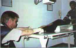
Illustration 4. Correct positioning of the dog for the diagnosis of the
dysplasia.
X-ray
perfeita:
to take place a x-ray of the articulations coxofemorais for the diagnosis of
the dysplasia, it is made neces-sária, preferentially, the general anesthesia,
could be of short duration, in such a way that the patient is free from any
reaction, with the objective of obtaining a correct positioning. The animal is
put then in number decubitus (Illustration 4), with the members extended
subsequent caudalmente, of equal length, parallel amongst themselves and in
relation to the spine, rotacionados mediante,de such form that the patelas are
put upon to the furrows trocleares. The pelve should be parallel to the
surface of the table, in other words, without inclination. For a x-ray of
appropriate positioning it is valuable a gutter, used to lie down the animal
in his/her interior, with the pelve out of the same. Therefore she is a very
important accessory for this exam type. The thoracic members are extended
cranialmente, becoming the care of there not being inclination of the thorax
of the animal. In these circumstances the image radiográfica should show us
the following (Illustration 5):
-
symmetrical ílios - oval pelvic channel, of symmetrical outlines, when
divided sagitalmente
-
forâmens symmetrical obturators
-
parallel femurs amongst themselves and with the spine - patelas put upon to
the furrows trocleares
The image radiográfica should allow the visualization of the whole pelve, as
well as of the articulations fêmoro-tíbio-patelares,para that she can evaluate
the symmetry of the ílios and the positionings of the patelas. If these have
not put upon to the furrows trocleares, was ended that the subsequent ones
were insufficient rotacionados or excessively. It is usually insufficient, in
other words, the patela tends the if to put upon more to the lateral côndilo
of the femur than to the furrow dito.No appropriate positioning of the
patelas, reached properly through the medium rotation of the members, a force
is exercised on the femoral heads, taking the dysplastic articulations to the
subluxação, while the normal animal won't run the same. It is usually this
subluxação the first alteration radiográfica and in beginning his/her more
importante.Através is that he/she is determined the degree in the index of
Norbeg. The other alterations will grow as a consequence of the subluxação, as
for instance the artrose, for that denominated of artrose secundária.Uma
quality x-ray should be well contrasted, being observed in a very detailed way
the board number acetabular and the head's structure trabecular and femoral
lap. They are reached these objectives been used good equipments of x-rays, it
is crans and films of good origin, revelation for processing automatic
whenever possible and a darkroom that it is really dark, provided of a lamp of
safety that is really of safety. Under the surface of the table radiográfica,
in Bucky, it is made present the grating anti-difusora, with the function of
absorbing most of the secondary radiation. This, when he/she is absent, it
produces images without contrast, that is, of filled with smoke aspect.
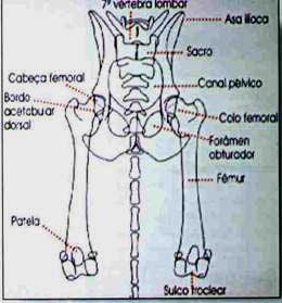
Illustration 5. I draw of the anatomical symmetry as consequence of a correct
positioning.
Inadequate x-ray:
it is that without appropriate positioning, characterized mainly by the
asymmetry of the ílios, au-sência of parallelism among the femurs, mainly for
abduction of the members, patelas no put upon to the furrows trocleares and
those without image pattern, for they be sub or super exposed (egg whites or
dark, respectively), harming the contrast, shaken, spotted, badly revealed,
etc., as well as those without data of the patient's identification in the
emulsão(antes of the revelação)do film.
Diagnosis:
accomplished through the index of Norberg (Illustration 6). He/she bases on
the determination of the centers of the femoral heads and of the union of the
same ones through a line, that will make possible to draw us, starting from
one of the centers a second line, that tangenciará the board acetabular
lateral skull. The two lines form an angle amongst themselves, called angle of
Norberg. This is just one of the necessary elements for the diagnosis of the
dysplasia. Other factors should be taken into account, such as the positioning
of the femoral head's center in relation to the board number acetabular, the
aspect of the line to articulate, the presence of alterations articulate
degenerative (secondary artrose) and the resignation of the boards
acetabulares, mainly of the lateral skull. According to Norberg the smallest
compatible angle with the normality is 105th, however it can have an
articulation with 105th or more and to be classified as close of the normal
(B) or slightly dysplastic (C), being Enough for this the osteófito presence
in the board acetabular lateral skull, adulterating the angle or when less
than 50% of the femoral head be inserted inside of the cavity acetabular. The
authors have been extolling at least 50%. It is of fundamental importance to
understand, that in beginning, as larger the angle of Norberg, adult will be
the consistency to articulate. In other words, adult will be the contact
between femoral head and cavity acetabular or adult will be the intimacy
between them or larger ser'ao fits of the femoral head. Starting from this
moment, as smaller the consistency to articulate, minor will be the angle and
more evident it will be the subluxação, could reach until the dislocation.
There are some years the Brazilian School of Veterinary Radiology - CBRV,
through a plêiade of doctors veterinary radiologists, has tornado reality, as
in other countries, the emission of a Certificate of Control of the Dysplasia
Canine Coxofemoral. This new services rendered modality appeared of a pressing
need, since there was an enormous discrepancy among the accomplished
diagnoses. These discrepancies took and they continue taking countless
creators to incommensurable damages, since they found his/her creation
supposedly in reproducers without dysplasia. CBRV, when receiving the x-ray
accomplished by veterinary doctor, he/she examines her as the diagnostic
quality, could return her, I marry the same doesn't obey to the demanded
technical patterns.
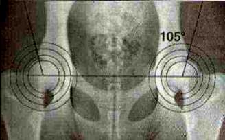
Illustration 6. The sobreposição of a concentric circumferences to the
femoral head's limit will determine the referred femoral head's center.
Norms of
CBRV for evaluation of the dysplasia coxofemoral in dogs in Brasil,segundo the
criteria of the Federation International Cinológica - FCI
1 - Technical
procedures
Age
The evaluation of the conditions articulate will be done conclusivamente
starting from the twelve complete months of age in most of the races,
exception made to Bullmastiff, Dogue of Bordeaux, Great Harms,
Leonberger,Maremma,Mastiff, Mastim Napolitan, Newfoundland,Landseer,Pyrenean
Mountain Dog and St. Bernard, whose appreciation should be accomplished with
at least eighteen complete months of age. Preliminary evaluations of the
articulations coxofemorais can be accomplished starting from the six months of
age.
Contention
With the purpose of assuring the wanted technical quality, it is obligatory
the patient's contention, by the use of pharmacological associations capable
to determine perfect relaxation of the animal, to obtain the correct
positioning and free from reactions on the part of the dog. The veterinary
doctor, when accomplishing the x-ray, will sign an affidavit, committing with
that contention type.
Positioning
Number decubitus with the pelvic members in extension caudal, parallel amongst
themselves and in relation to the column vertebral,tomando-if taken care of
maintaining the articulations fêmoro-tíbio - patelares rotacionadas
medialmente, in such a way that the patelas are put upon to the furrows
trocleares.Deve-if still to be the careful for the pelve to be in horizontal
position. A second x-ray will still be able to be used, with the pelvic
members inflect-frog position (frog position).
Identification of the film
In the permanent minimum identification of the film, in his/her emulsão,deverá
to consist the number of registration of the animal, race, date of birth, it
dates from the exam radiográfico and the identification of the articulation
coxofemoral right or left.
The patient's identification
The
veterinary doctor when accomplishing the x-ray should identify the animal,
case is not still, for microchip, correctly denominated of transponder(Figura
7), or for tattoo, for a subsequent control, if necessary.
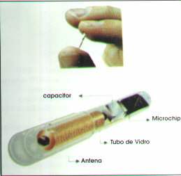
Illustration 7. The transponder (microchip) it measures 11 x 2mm. His/her
implantation
is subcutaneous, as any administration medicamentosa for the same road,
dorsalmente to the encounter of the scapulas.
Size of the film
It
should be enough to include the whole pelve and the patient's articulations
fêmoro-tíbio-patelares.
Quality of the x-ray
The
x-rays will be analyzed properly identified and the ones that obey the
criteria of positioning of the animal, whose quality pattern offers conditions
of visualization of the head's personal computer bone trabeculação and femoral
lap and still definition needs the margins of the articulation coxofemoral,
especially of the board number acetabular.
2 – Laud
The
radiologist, when receiving the x-ray, appraised his/her quality for the
diagnosis, being to his/her position the possibility of being returned to the
veterinary doctor that accomplished her, in case it doesn't obey the wanted
technical patterns. For the emission of the definitive decision, each x-ray
will be examined by one of the radiologists accredited by CBRV, chosen for
draw, that he/she won't have knowledge of the registration name or even of the
proprietor of the animal. Each proprietor will be entitled, by payment of the
respective costs, of appealing to one second and last diagnosis, submitted to
the jury of the Displasia Coxofemoral of the Scientific committee of the
Federation International Cinológica.
Classification of the articulations coxofemorais:
A (HD -):
without signs of dysplasia coxofemoral (Illustration 8)
The
femoral head and the acetábulo are appropriate. The board acetabular lateral
skull comes sharp and lightly round. The space to articulate it is narrow and
regular. The angle acetabular, according to Norberg, is of aproximadamente105º,
as reference.
B (HD
+/-):
articulations
close coxofemorais of the normal (Illustration 9)
The femoral head and the acetábulo are lightly incongruous and the angle
acetabular, according to Norberg, is of approximately 105th or the femoral
head's center comes medialmente to the board number acetabular.
C (HD +):
dysplasia light coxofemoral (Illustration 10)
The femoral head and the acetábulo are incongruous. The angle acetabular,
according to Norberg, is of approximately 100th and/or there are a quick
flattenings of the board acetabular lateral skull. They can be present
irregularities or just small signs of alterations osteoartrósicas of the
margin cranial acetabular, flow or number or in the femoral head.
D (HD ++):
dysplasia moderate coxofemoral (Illustration 11)
Evident incongruity between femoral head and the acetábulo with subluxação.
Angle acetabular, according to Noreberg, is larger than 90th, as reference.
Presence of flattening of the board acetabular lateral skull and/or signs
osteoartrósicos.
E (HD +++):
displasia severe coxofemoral (Illustration 12)
Marked dysplastic alterations of the articulations coxofemorais, as
dislocation or different subluxação. Angle acetabular, according to Norberg,
smaller than 90th. Evident flattening of the margin cranial acetabular,
deformation of the femoral head (mushroom format, flat) or other osteoartrose
signs.
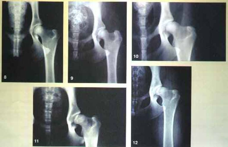
Illustration 8. The (HD -), without signs of dysplasia
coxofemoral.
Illustration 9. B (HD + / -), articulation close coxofemoral of the normal.
Illustration 10.C (HD +),displasia coxofemoral leve.Discreta subluxação.
Illustration 11.D (HD ++),displasia moderate coxofemoral. Evident subluxação,
accompanied of osteoartrose.
Illustration 12.E (HD +++), dysplasia severe coxofemoral. Subluxação still more
evident, accompanied of osteoartrose.
Pré requesitos for the emission of the decision of dysplasia coxofemoral for
CBRV:
-
X-ray of the articulations coxofemorais according to the norms of CBRV.
-
Authenticated copy of the pedigree or of the stripe of the animal.
-
The veterinary doctor's affidavit *
-
The proprietor's affidavit or responsible *
-
It
rates in money or order check to ABRV
-
All of the x-rays directed to CBRV should be sent of any part of Brazil for:
Brazilian school of Veterinary Radiology
Post office box 42041 - 04073-970 - São Paulo - SP
PHONE: (0_11) 530-9050
-
*
The affidavits should be requested CBRV
Treatment:
it can be medicamentoso or cirúrgico.Relacionam-if in this last several
possibilities, from the simplest, such as, for instance, the pectineotomia and
femoral head's ressecção (artroplastia excisional), until the most complex, as
the corrections of deviations of the type valgus geno and antiversão, the
triple osteotomia of pelve, the osteotomia intertrocantérica, the prolongation
of femoral lap, the total prosthesis, etc., and the surgical associations, as
femoral. Modernamente has if treaty, not only the dysplasia coxofemoral, but
also the dysplasia of the elbow, the osteocondrose, head's femoral,a
espondiloartrose avascular necrosis, finally, all of the pathologies
articulate inflammatory degenerativas(artroses)e (arthritides) through
products of natural origin with the property of regenerating (anabolizar)e to
protect the cartilage to articulate degenerate, producing a natural analgesia.
The steroidal anti-inflammatories chewed the pain, liberating the movements
articulares.Estes steroids added to the movements articulate has a destruction
action (catabolização)da cartilage to articulate, that it is antagonistic to
the factors anabolizantes of the products above referred. For this reason the
association of the same ones should not be recommended, much less only the
application of the anti-inflammatories. The action anabolizante of the product
can have a better when accompanied of appropriate measures of manejo,tais
final result how to maintain the animal in restricted places so that the same
reduces his/her physical activity, as well as avoiding the patient's obesity
and the slippery places. There is the possibility to happen a remodelamento
osteoarticular besides. This fact is of addition importance, because the
osteófitos pericondrais could be, at least, partially reabsorbed,
decompressed, for instance, the ramifications nervous located eferentes in the
spaces intervertebrais. We could avoid the calcification of the disks
interverterbrais.
In case these procedures are not crowned of success, we cannot stop
considering the surgical intervention as an additional possibility.
Bibliographical
references
1 - BRAUND, K.G. Hip dysplasia and degenerative myelopathy:marking the
distinction in dogs.Veterináry Medicine, (aug.),1987.
2 - CORLEY,E.A.;KELLER,G.G.Hip dysplasia:a guide for dog breeders and owners.
2nd. edition, Orthopedic Foundation for Animals,1989.
3 - DOUGLAS, S.W.; WILLIANSON, H.D.Veterinary radiological
interpretation.Philadelphia,Lea & Febiger,1970.
4 - ETTINGER,S.J.Textbook of veteranary internal medicine.Philadelphia,W.B.
Saunders,p.2211-14,1983.
5 - FOX,S.M.;BURT, J. Symposium on hip dysplasia.Veterinary
Medicine.p.684-716,1987.
6 - KEALY,J.K. Diagnostic radiology of the dog and cat.Philadelphia, W.B.
Sanders,1987,p.352-362.
7 - MORGAN, J.P.; STEPHES, M. Radiographic diagnosis and control of canine
dysplasia. Iowa State University Press, 1988.
8 - SMITH, G.K. et al. New concepts of coxofemoral joint stability and the
development of a clinical stress-radiographic method for quantitating hip joint
laxity in the dog. Journal of American Veterinary Medical Association,
v.196,n.1, p.59-70,1990.
9 - THRALL, D.W.;LEBEL, J.L.Carlson's veterinary radiology. Philadelphia, Lea &
Febiger,1977.
10 - TOMILINSON,J.; McLAUGHLIN. Jr., R.Canine hip dysplasia: developmental
factors, clinical signs and initial examination steps.Veterinary Medicine,
p.25-53,1996.
11 - TOMILINSON,J.; McLAUGHLIN. Jr., R.Medically managing canine hip
dysplasia.Veterinary Medicine, p.48-53,1996.
12 - TOMILINSON,J.; McLAUGHLIN. Jr., R.Total hip replacement: the best treatment
for dysplasia.Veterinary Medicine, p.118-143,1996.
13 - TOMILINSON,J.; McLAUGHLIN. Jr., R.Symposium on canine hip dysplasia.
Veterinary Medicine, p.25-23,1996.
14 - TOMILINSON,J.; McLAUGHLIN.
Jr., R.Actualidad en displasia coxofemoral. El perro ovejero alemam,p.41-43,1997.
15 - VERLAG, M.; SCHAPERH, H. Bercht der hüftgelenk dysplasia.
Kleintier Praxis, n.23,p.169-180,1978.
Edgar
Luiz Sommer - CRMV-SP nº 1556 1. Sócio proprietário do Provet, responsável pelos
setores de radiologia, ultra-sonografia e ecocardiografia; Conselheiro do
Conselho Regional de Medicina Veterinária do Estado de São Paulo; Diretor
Secretário do Colégio Brasileiro de Radiologia Veterinária; Diretor pela América
do Sul do International Veterinary Radiology Association.
Carlo
Leonardo Grieco Fratocchi - CRMV-SP nº 7080 2. Presidente da Associação
Brasileira de Radiologia Veterinária; membro do Colégio Brasileiro de Radiologia
Veterinária; Radiologista do Provet; membro do International Veterinary
Radiology Association.
Fonte deste artigo: Revista
de Educação Continuada do CRMV-SP.
São Paulo, fascículo 1, volume 1, p.031-035, 1998.
Edgar Luiz Sommer - CRMV -
SP 1556
Carlo Leonardo Grieco Fratocchi - CRMV - SP 708
Provet
Av. Aratãs, 1009
Cep: 04081-004 - São Paulo
SP -Brasil
provet@uol.com.br