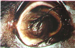
Illustration 1 - dog Eye presenting dermoid corneal. To notice cutaneous formation and their enclosures in cornea.
CORNEAL DISEASES IN SMALL ANIMALS
Anatomofisiologia of the cornea
The cornea is a transparent disk to circulate and concave-convex that it occupies the portion rostral of the obo(3,4). It consists of a singular arrangement of conjunctive fabric, cells endoteliais and membranes basal, avascular and transparent, that he/she has for functions: to transmit and to refract the light, for an ideal vision and to serve as a protecting barrier for the contents ocular internos(22,27). The cornea of the dog is composed by five layers pré-corneal tear distintas:filme, epithelium, estroma, membrane of Descemet and endotélio (25). It is capable to transmit the light because of the regular arrangement of the collagen in the estroma(27), of the lack of pigments, vases and of the maintenance of a relative desidratação(22 state). It is worth to stand out that the maintenance of such a state of corneal deturgescência, so important for the transparency of the same, it is guaranteed by a dependent mechanism of energy (bomb of NA+ K+ ATPase)presente in great amounts in the endotélio and in the epitélio(23,25).
The corneal epithelium is of the type stratified pavimentoso no queratinizado and he/she understands several layers of polyhedral cells anchored on a basal membrane, that it promotes the adhesion of the epithelium to the adjacent estroma. The estroma is formed by fine fibers colágenas of the type I and II, disposed in form of parallel plates and for queratócitos, responsible for the formation and maintenance of the fibrous lamellae, constituted more than 90% of the corneal substance. Linfócitos,macrófagos,neutrófilos,assim as some enzymes are present.
The membrane of Descemet is fine, firm, acetabular, elastic and seemingly homogeneous. To the electronic microscopy two layers are observed poorly defined. One of them, adjacent to the estroma, it consists of collagen type II and he/she has his/her thickness increased with the age. The other is the modification of the membrane of the endotélio.
The endotélio possesses a single layer of flat cells that you/they are in the subsequent portion to the flat ones that you/they are in the subsequent portion to the membrane of Descemet. The nutrition of the cornea is obtained through the tear film, diffusion of metabólitos of the vasculatura perilímbica and internally for the humor aquoso(23). The removal of the catabolic residues happens for the same vias(3).
The cornea is supplied richly by derived sensorial nerves of the division of the equal of cranial nerves fifth. The nervous trunks penetrate in the estroma close to the limbo, advancing radialmente towards the central cornea, where they ramify repeated times, finishing in the epithelium as endings nervous livres(3).
Mechanisms involved with the corneal repairing
After an offense that damages or it removes the corneal epithelium, the cicatrization of the lesion follows a systematized sequence of even-tos (15). Elapsed a hour of the offense, the cells of the basal layer of the epithelium begin the if it smoothes and six hours after the lesion, these, for sliding, collect the corneal defect. The mitosis is active three to four days after the offense and at the end of 5 to 7 days the cornea comes íntegra(3,4,15 seemingly).
Defects involving the epithelium and the previous estroma also heals for the sliding epitelial and mitosis (1). For his/her relative inactive metabolic state, the estroma develops for repairing of the type cicatricial in a slower way in face of the complexity that involves the process reparatório(15). Lesions estromais no complicated they can be scarred in an avascular way; however, in infected lesions or destructive, the presence of vases is a constant, as well as it happens at other places of the corpo(15).
The ulcers become complicated when there are persistence of the cause, adherence and bacterial proliferation, parallel to the action of enzymes líticas(3).
Semiology of the cornea
Ocular exam and diagnostic tests complemental
The exam oftalmológico should include a systematic evaluation of all the ocular structures, beginning with the enclosures and millstone-blindfold-if in felt antero-posterior(15).
Numerous diseases can be found in the margins palpebrais, including trichiasis, eyelash ectópico, nasal pleats, distriquíase,tumorações, traumatic lesions, entrópio and ectrópio. The function of the eyelids is evaluated by the frequency of his/her sincursões (being considered as normal four to five incursions per minute) and the extension of the closing palpebral (15). The tear production is evaluated, through the test of the tear of Shirmer(3). According to PERUCCIO et al. (20), the normal values for the dog vary between 15 and 25mm per minute; smaller values than 10mm per minute are suggestive of deficiency in the tear production. The cat presents normal values discreetly inferior and more variables. Drugs parassimpatolíticas and local anesthetics reduce the tear production. In spite of, the manipulation of the eye and the use of the fluoresceína can increase these valores(25).
The corneal sensibility should be evaluated playing one sweats surface. Continuous action is instilled anesthetic eye drops (proparacaína hidrocloridrato to 0,5%) in the sack conjutival for, soon afterwards, with a small forceps, to expose the medial face of the third eyelid and, examining her/it in his/her surface, to locate strange bodies, woven hiperplásicos, inflammation and folículos(9 formation).
The good exam of the cornea forces to use the biomicrocópio in rift lamp, though such equipamento,face his/her cost, is not to the reach of most of the proficionais. Alternatively, the exam can be driven with magnifying glass Peak and a source of artificial light, as the transiluminador of Finoff(3).
The cornea ulcers cannot be visible clearly, even with a good illumination; for this reason, all of the suspicious eyes should receive the test of the fluoresceína(25). The external dyeing is useful in the diagnosis of corneal lesions, since the intact epithelium, for his/her high lipidic content, hinders the penetration of the hydrophilic color not being for him colored. Any rotura in the barrier epitelial will allow the fast penetration of the fluoresceína in the estroma and his/her fixation. ( 1).
The job of the Pink color of Bengal is less admitted, however it is useful in the diagnosis of the ceratoconjutivite seca(15). This test allows to check the degree of deterioration of the cells epiteliais and to detect erosions intra-epiteliais dendritic caused by herpesvirus, that are of difficult detection for the fluoresceína(25,12,20).
Swabs and scraped conjutivais should be extolled for the isolation and identification of eventual pollutants.
Corneal reactions to the ceratopatias
The largest barriers to the cornea edema are the endotélio and the complete epithelium. When one of them is damaged, there is soak of the estroma. Ofenômeno can be related the a variety of causas:distrofia endotelial,dano endotelial associated to the membrane persistent pupilar, trauma poisonous mecânico,reações, uveitis anterior,ndotelites,glaucoma,neovascularização and superficial ulcerations or normal horny profundas(26)A doesn't contain blood vessels. The vases, that can be superficial or deep, invade the estroma in in answers to the injuria(25).
In the deep vascularização, these appear under the margin limbic escleral, with little tendency to the ramificação:são darkened by the projection of the esclera in the limbo. The vases superficias can be seen of I permeate to the limbo and originally they are conjutivais (2,23).
When the neovascularização retreats, such vases lose his/her content intraluminal, however, their walls stay. The theses feel the name of" vases fantasmas"que can be visualized as pale lines in the cornea for the retroiluminação(2).
Lesions complicated estromais, especially when the offense persists, it maintains the vases neoformados and they propitiate the genesis of the granulation fabric.
The repairing of the cornea is accomplished by the fixation of keratocytes and invasion of fibroblasts and macrophages. Fibers of collagen produced are deposited hindering the passage of the light irregularly.
The pigmentation results of the migration of coming melanócitos of the limbo and of woven perilímbicos, being more commonly associated to the chronic inflammation. According to GELATT(6), the corneal pigmentation is found in disorders as the chronic superficial keratitis (panus), syndrome of the pigmentary keratitis in races braquicefálicas, ceratoconjuntivite dries and chronic ulcerative keratitis. The pigmentation can also happen in the chronic glaucoma and, in this case, to come accompanied by degenerative corneal changes.
Main abnormalities of the development and congenital diseases
The microcórnea is usually associated to the microftalmia. It is hereditary anomaly and of occurrence in the races Poodle, Sch-nauzer miniature and Collies. The clinical sign corresponds her/it a cornea of inferior diameter to 12 mm, accompanied of increase of the portion escleral,sem exoftalmia (26).
The term megalocórnea refers to a cornea of larger size than his/her normal one and of horizontal diameter of approximately 16 to 18 mm. It is a congenital anomaly, rare in dogs and usually associated to the congenital glaucoma and to the buftalmia(26).
The dermoid is a mass of cutaneous fabric that he/she appears in a position ectópica. It is usually found in the temporary area of the limbo, extending to the cornea and the esclera. These formations contain epithelium queratinizado, hair, blood vessels, fibrous fabric, fat, nerve fibers, glands, flat musculature and they can still contain cartilage (Illustration 1). His/her removal forces to accomplish procedures in keratectomy superficial(26).
Illustration 1 - dog Eye presenting dermoid corneal. To notice cutaneous
formation and their enclosures in cornea.
Dystrophies and corneal degenerations
The degenerations and corneal dystrophies exhibit similar characteristic, but they represent different pathological processes. The degenerations are the result of local inflammations and they can be associated with systemic disorders, as well as with senile processes. The dystrophies are disorders of the development, frequently hereditary, that you/they affect the axial cornea harming the vision. According to the layers of the cornea attacked, the dystrophies can be subdivided in: epiteliais, estromais (previous and subsequent) and endoteliais(6).
Dystrophy epitelial
The dystrophy epitelial is probably the responsible for the syndrome of the difficult corneal erosion. The erosions difficult epiteliais, persistent úl-waxes, appealing erosion, indolent ulcers and ulcers of Boxer are denominations for the superficial ulcers, that you/they tend to the recorrência(6). In the difficult ulcers the lesions are due to the separation between the corneal epithelium and the estroma, probably for defects in hemidesmossomos juncionais between the basal cells of the epithelium and their membrane basal(25). This condition was described originally in dogs of the race Boxer and, probably, he/she feels in other races (8).
The diagnosis is based in the clinical signs, in them history and in the ophthalmic semiotécnica. Many animals come, initially, with sharp ocular pain, evidenced by fotofobia and blefaroespasmo. The ulcers happen spontaneously without any history of previous trauma plows characterized by his/her chronic course, superficial nature, vascularização speech and of other inflammatory signs. The edema can be discreet the moderate and the borders of the ulcers usually meet irregular face to the loss epitelial circunjacente (4,19). The visual therapy goes the difficult ulcers consists of the debridamento of the epithelium in the adhered, with or without chemical agents. The removal of this favors the multiplication of adjacent epithelium and the production of the basal neomembrana and hemidesmossomos(26). The topical antibiotics of wide spectrum plows suitable in the prophylaxis of bacterial infections. The atropine to 1% topical it i admitted the marks of softening the pain produced by the uveitis reflexa(26).
Dystrophies and degenerations estromais
The corneal lipidose i the frequent degeneration in the horny of senior dogs. In the German Shepherds it has been associated to the hiper-colesterolemia and hipertrigliceridemia(6). The lipidose also shows the the bilateral corneal dystrophy in Poodles toy and young miniatures, in Afghan Hounds and in other races. White deposits plows evidenced, of translucentes the opaque ones, usually located in the previous corneal estroma. The neovascularização, in these marry, can be presente(6).
There plows marry that these deposits motivate the occurrence of superficial corneal inflammation severe. If the lipidose to interferes in the vision or to allow that there i discomfort, the superficial keratectomy i suitable, seeking the removal integral or partial of the lesion. Appeals powders keratectomy plows habitually infreqüentes(6).
Dystrophy endotelial
The dystrophies endoteliais have been described in the Boston terrier, Dachshund and Chihuahua. The condition usually attacks the senior dogs and he/she shows with signs of edema of slow progression initially in horny axial the. Other signs oculars plows in general inexistent. With the progression, they can happen vascularização, pigmentation and keratitis bolhosa(6).
The cornea edema can be minimized by the instilação of hypertonic agents the the chloride of sodium in variable concentrations from 2 to 5%, the well goes them the glucose job in hypertonic solution. In spite of the benefits obtained with these solutions, adds patient ones can become uncomfortable face to the irritation goes them produced. In these marry it i recommended to interrupt his/her emprego(6).
Neoplasias
The neoplasias of horny the plows rare almost always represent the secondary extension of lesions with primary ranch in another segment of the ocular bulb. The melanomas epibulbares or limbic they usually eats in the form of pigmented lesions; though there plows marry in that you/they plows of variable progression and they develop slowly in dogs idosos(6,26).
Adenocarcinomas, the well the the carcinomas of scaly cells, rarely happen in the horny of dogs. They eats of white and rosy coloration, the high masses and multibuladas. In most of the marry, there i in the involvement of the limbo(26). The treatment consists of the local excisão of the mass, goes combined superficial keratectomy with procedures in crioterapia or Beta radiation, seeking to reduce the recorrência(6,26).
Keratitides
Keratitides superficial punctata
The keratitides punctatas can eats united or bilaterally the superficial defects of the corneal epithelium. The center of the scan-sões puntiformes i colored with fluoresceína. It adds signs observed clinical são:conjutivite, blefarospasmo and epífora. The vascularização of the horny can happen in marry crônicos(13).
This pathology can be induced by countless conditions aviltantestais the: chronic exhibition of the horny in patients under effect of general anesthesia, exhibition keratitides in races branquicefálicas and the job of anesthetics tópicos(25). The specific therapy, since the causal agent's determination, it can be dificultada(25).
"Flowered Spots"
Corneal opacities in animals that inhabit tropical and subtropical climates were described recently in the dog and in the cat. The lesions, that passed to Florida Spots" to be denominated", they plows characterized by opacificações of coloration white or white-grayish, of sizes variados,circunscritas and multifocais, found in the estroma corneano(Figura 2). It i condition in the responsive to the corticoterapia, to the antibioticoterapia the well the to the topical antimicóticos. The cornea eats without any inflammation indications and the eyes don't exhibit any discomfort or irritation. The function or functional vision i rarely afetada(26).
Chronic superficial keratitis (Panus)
The chronic superficial keratitis i an illness that happens dogs, of progressive and inflammatory character, could lead to the ce-gueira. The synonymies, the terms plows recognized: Panus of the German Shepherd, degenerative Panus, Keratitis superficial estromal and Syndrome of Uberreiter's(26).
The condition shows bilaterally in the form of lesion red, vascularized, with infiltration subepitelial of conjunctive fabric. Usually the corneal epithelium stays intact, and the migration of pigments (corneal melanosis) commonly it accompanies him infiltrated inflammatory fibrovascular, that it invade the estroma anterior(26).
Relatively his/her aetiology, face to the therapeutic answer the imunossupressivos, i believed to treat of illness imunomediada(24,13,6,26). The disorder presents, still, positive correlation with high altitudes and increase of the levels of ultraviolet radiation. The lot of sào the races that can develop Panus; in spite of, he/she i the German Shepherd the acometido(24 lives). The diagnosis i based in the symptoms, that exhibit bilateral lesions composed primarily by vascularização, pigmentation or goes the combination of both (Illustration 3). The lesions involve the temporary corneal quadrant in lives than 95% of the casos(24).
The treatment consists of the uses of topical anti-inflammatory agents potent the the corticosteroids or the cyclosporin A. In the marry of very extensive lesions and in the responsive to the clinical therapy, superficial keratectomy or the application of Beta radiation have been per teams preconizada(24). It i recommended the therapeutic alternative, the cryosurgery, using the nitrogen liquidates or rust nitroso(24).
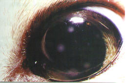
Illustration 2 - dog Eye with" Florida Spots." Opacity corneal
puntiforme i observed, bounded and multifocal.
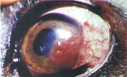
Illustration 3 - dog Eye evidencing the chronic superficial
keratitis (Pannus).
To notice conjunctive fabric richly vascularized invading horny the.
Pigmentary keratitis
The pigmentary keratitis i common condition in dogs in that the pigment i carried to the epithelium and superficial estroma, in association with keratitides crônicas(25). The pigmentation ranch in the horny can allow the identification of the irritation source. The focal pigmentation i habitually associated to the distiquíase, entrópio, ectrópio, nasal pleats and eyelashes aberrantes(6). The central pigmentary keratitis happens in the ceratoconjutivite dries, keratitis goes exhibition, lagoftalmia and keratitis neuroparalítica(6).
The treatment consists of the removal of the agent causal(6). Topical Corticoperapia (in the absence of ulcers), preparations with artificial tears, stimulation of the loom production goes the uses of oral pilocarpina or of the topical cyclosporin, the well the procedures in superficial certactomia can be useful in adds casos(6,25).
Ulcerative keratitis
The ulceration i the corneal disease lives commonly observed in dogs and cats and it i characterized by the loss of the epithelium and of the estrus-ma superficial, with or without loss of the fabric cornenano profundo(13,5).
The ulceration of the horny exhibits wide causas,mas variety the trauma i, probably, the most common among the agents envolvidos(10).
The infectious cause include: bacterial infections (possibly preceded by an initial trauma) it goes Staphilococcus and Pseudomonas aeruginosa, infections micóticas the the aspergilose and the candidiasis and viral infections goes the Herpesvirus feline type I (10,13,23).
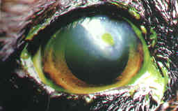
Illustration 4 - dog Eye
presenting corneal ulceration.
To notice depression and edema perilesional
The ulcers can be secondary to other corneal illnesses, the the dystrophies epiteliais, the chronic edema in the marry of glaucoma and the keratitides neurotróficas(25). When to the substance loss the strange bodies have been incriminated, abnormalities of the eyelashes (distiquíases, trichiases and eyelashes ectópicos), scratch goes cat, chemical traumas (acids and álcales), abnormalities palpebrais(entrópio, ectrópio, lagoftalmia or exoftalmia and buftalmia). There i, still, cause related to the paralysis of the facial nerve and diseases of the film lacrimal(10,12,4,2,15,25,5).
The ulcers frequently exhibit classic clinical signs, translated by fotofobia, bleforospasmo, epífora and loss of the transparency goes the invasion of vases, migration of inflammatory cells due to the edema, disarrangement of the lamellae of collagen, resulting from the repairing cicatricial, deposition of pigments and of other substances the lipids and cálcio(25,22). The superficial ulcers plows usually small and resultants of offense mecânica(4). The deep ulcers plows usually of forms ovuladas or round, of abrupt margin and surrounded by edema and infiltration vascular(17)(Figura 4). Among the complications, they stand out the perforations and the uveitides secundárias(25).
The therapy consists of the prophylaxis or in the control of the infection, of the erosion and in the retreat of the causa(13). In general the, the treatment should be addressed goes the prevention or elimination of the contamination, control of the uveitis, analgesia, interruption of the destruction tecidual, preservation of the transparency and of the corneal function and support tecidual(10).
Herpetic keratitis
Viral keratitides causes them of ulcerative keratitides in dogs have been incriminated, however there i in the comprovação(10). To the con-trário of the dogs, in cats, the feline Herpesvirus i important causes of ulceration corneana(9). the herpetic keratitis can attack felines of any acts group; though they plows recognized lives them frequent the marry attacking adult animals with light signs of afecção of the superior breathing treatment. He/she fits to remind that it i treated of afecção manifests united or bilateralmente(16).
The herpetic ulcers eats second an or several patterns. They can be small and numerous (punctatas), lineal and ramified (dendritic ulcers) and geográficas(19). The clinical signs still exhibit conjunctivitis of light the moderate ones to the perforation of the horny with loss of the affected eye (16). Topical antiviral agents should be used goes, in the minimum, two weeks. The trifluridina, drugs of choice, it i particularly suitable, since it penetrates the córnea(7 better). In our half, the idoxuridina have also been used.
Ulcerative keratitis with horny highjacking in felines
The chronic ulcerative keratitis, with highjacking of the estroma corneano,é of exclusively occurrence in the felino.A condition i still of-nominada of focal mummification or keratitis necrosante (14,7). In the corneal kidnapping the lesion can eats in the plate form, high or superficial, central or paracentral and well bounded, oval and of coloration chestnut-dark or black, accompanied by epífora, blefarospasmo, opacity, hiperemia conjutival and, occasionally, quemose(27,7).
Regarding the countless incursions accomplished in the sense of establishing the causes, it i i not known still. caustic agents, malformation palpebrais, ceratoconjutivite dries, feline herpesvirus, trauma and bacterial infections have been included among the probable precursors of the corneal kidnapping (14,7). The treatment consists of the excisão of the focal lesion goes the superficial keratectomy. Antibioticoterapia profilática and drugs ciclopégicas can be associated. In the deep lesions the job of grafts or of conjutiva pedículos it can be indicado(7).
Ceratoconjutivite dries / CCS/KCS
The ceratoconjutivite dries or dry eye i common ophthalmic problem in dogs. The condition usually results of the deficiency of the aqueous component of the pré-corneal loom film and it exhibits several facets: racial predisposition, hypothyroidism, paralysis of the facial nerve, medicines (atropine, sulfonamidas), surgical excisão of the gland of the third eyelid, conjunctivitis and cinomose have been incriminados(28). Recent studies have been showing that the much KCS of the dogs the the one of individuals of the human species they have his/her genesis starting from alterations of the system imunogênico(18).
The diagnosis i based in the clinical signs and in the results obtained with loom test of Schirmer. The outstanding sign in attacked patients translates himself goes mucoid ocular secretion the mucus-festering, that he/she adheres to the epithelium and that, usually, it accompanies loss shine in the horny and hiperemia conjuntival(28) (Illustration 5). Sharp marry can produce superficial ulcerations, deep and until the perforation of horny the. Though they plows found chronic superficial manifestations with progressive deterioration of the vision. The vascularização and the pigmentation plows of occurrence sistemática(28).
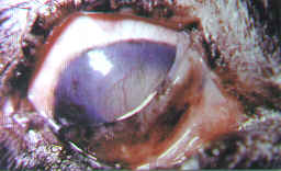
Illustration 5 - I Look of the dog presenting dry ceratoconjuntivite.
Be noticed edema, neovasos, deposits of pigments and discharges ocular
periocular and.
The conventional therapeutic approach consists of frequent instilações of artificial loom, drugs anti-inflammatory, mucolytic and antibiotics; and of oral the uses of pilocarpina - ophthalmic solution to 1%, instilled 1 to 2 drops the day, in the main meal. Now the cyclosporin THE, in the eye drops form or ointment, in different concentrations and to intervals of 12 or 24 horss, it have been used in the relief of the clinical signs and in the lacrimogênese. ( 21,25,28).
BIBLIOGRAPHY
1. BISTNER,G.A. et al. Surgery of the cornea. In: Athas of veterinary ophtalmic sugery. Philadelphia, W.B.Saunders, 1977, p. 157-79.
2.
BLOGG,G.R.Disease of the cornea. In: The eye in veterinary practice.
Philadelphia, W.B.Saunders, 1980, p.374-424.
3 –COL
3. LINS, W.W.; RENDA, J.A.Olho e ouvido. In:THONSON, R. G. Patologia veterinária especial. São Paulo, Manole, 1996, cap 127, p. 1086-92.
4. DICE, P.F. The canine cornea. In: GELATT,K. N. Veterinary ophthalmology. Philadelphia, Lea e Febiger, 1981, p. 343-73.
5. DZIEZYC, J. Ulcerative Keratitis. In: KIRK, R.W. Current veterinary therapy XII. Philadelphia, W. B. Saunders, 1989, p. 656-8.
6. GELATT, K. N. Corneal diseases in the dog. In: GLAZE, M. B. The compendium collection::ophthalmology in Small animal practice. 2. ed. New Jersey, Veterinary Learning Systems. 1996, p. 107-13.
7. GELATT, K. N. Feline ophtalmology. In: GLAZE, M. B. The compendium collection: ophthalmology in small animal practice. 2. ed. New Jersey, Veterinary Learning Systems. 1996, p. 201-9.
8.
GELATT,K. N.; SAMUELSOM, D.A. Recurrent corneal erosions and epithelial
dystrophy in the boxer. Journal of the American Animal Hospital Association.
v.18, p. 453-60, 1982.
9 -KERN, T.J. Diseases of the cornea and sclera. In: BIRCHARD, S.J. e SHERDING,
R. G. Small animal practice. philadelphia, W. B. Saunders, 1994, vol.1,p.
1197-207.
9. KERN, T.J. Ulcerative Keratitis. Veterinary Clinics of North America:Small Animal Practice. V.20,n.3, p.646-66, 1990.
10. KIRSCHNER,S.E. et al. Diseases of the cornea and sclera. In: MORGAN, R. V. Handbook of small animal practice. Philadelphia, W. B. Saunders, 1992,p. 1063-76.
11.
LAFORG,H. Diagnóstico y tratamiento de las úlceras corneales.
Waltham International Focus. v. 3,n. 1,p. 2-8, 1993.
13 -MAGRANE, W.G. Canine ophthalmology. 3. ed. Philadelphia, W. B. Saunders,
1977. p. 107-44:: Diseases and surgery of the cornea and sclera.14 -MORGAM, R.V.
Feline corneal sequestration: a retropesctive study of 42 cases ( 1987 - 1991 )
Journal of the American Animal Hospital Association. V.30,p.24-28, 1994.
12. NASISSE,M.P. Canine ulcerative Keratitis In: GLAZE,M.B. The compendium collection::ophthalmology in small animal practice. 2. ed. New Jersey, Veterinary Learning Systems, 1996. p.45-57.16 -NASISSE,M.P.Manifestations diagnoses, and treatment of ocular herpesvirus infection in the cat. Continuing Education v.4, n.12, p.962-968, 1982.
13. NELSON,D.L.;MACMILLAM,A.D.Doenças da córnea. In: KIRK,R.W. Atualização terapêuticva veterinária. São Paulo, Manole, 1988,p. 808-17.18 -OLIVERO,D.K. Clinical evaluation of 1% cyclosporine for topical treatment of Keratoconjuntivitis sicca in dogs. Journal of the American Veterinary Medical Association. V.199, n. 8, p.1039-46, 1991.
14. PENTLARGE,V.W. External pphthalmic disease and glaucoma In: LORENZ, M. D. et al. Small animal medical therapeutics. Philadelphia, Lippincott 1992, p. 389-456.
15. PERUCCIO,C. et al. Diagostics. In: PEIFFER,R.L.;PETERSEN-JHONES,S. M. Small animal ophthalmology. 2. ed. Philadelphia, W. B. Saunders, 1997, p. 1-12.
16. PETERSEN-JONES, S. Ocular discharge. In: PEIFFER,R.L.;PETERSEN-JONES,S.Small animal ophthalmology. 2. ed. Philadelphia, W.B.Saunders, 1997,p. 197-225.22 -RENWICK,P. Diagnosis and treatment of corneal disorders in dogs. In Practice v. 18, n.7,p. 315-28, 1996.
17. RENWICK,P;PETERSEN-JONES,S Orbital and ocular pain. In: PEIFFER,R.L.; PETERSEN-JONES,S.Small animal ophthalmology. 2.ed. Philadelphia, W.B.Saunders, 1997,p. 167-196.
18. ROBERTS,S.M. Pannus. In: KIRK,R.W. Current veterinary therapy XII. Philadelphia,W.B.Saunders,1995,
19. SLATTER, D. Fundamentals of veterinary ophthalmology. 2.ed. Philadelphia,W.B.Saunders, 1990.p.257-303:
20. Cornea and sclera.26 -WHITLEY,R.D. Veterinary ophthalmology. 2.ed. Philadelphia, Lea e Febiger,1981, Cap9, p. 307-354: Canine cornea.
21. -WILKIE, D.A.Diseases and surgery of the eye. In: SHERDING, R.G. The cat diseases and clinical management. 2.ed. New York, Churchill Livingstone, 1994,vol.p.2011-2046.
22. WILKIE,D.A.Management of Keratoconjutivitis sicca in dogs. In: GLAZE,M.B. The compendium collection:ophthalmology in small animal practice. 2.ed. New Jersey, Veterinary Learning Systems. 1996.p.234-238.
José Luiz Laus 1 -
CRMV-SP nº 3375,
Prof. Adj. Dept. Clínica e Cirurgia Veterinária da
FCAV - UNESP/ JABOTICABAL
e-mail: jllaus@fcav.unesp.br
Arianne Pontes Oriá 2 - CRMV-SP nº 1834,
Médica Veterinária, Estagiária do Serviço de Oftalmologia
do Hospital Veterinário
"Governador Laudo Natel" da
FCAV - UNESP/ JABOTICABAL
* Extraído
de: Revista de Educação Continuada do CRMV-SP / Continuous Education Journal
CRMV-SP
São Paulo, volume 2, fascículo 1. p26 - 33, 1999.
* Com Autorização de Prof. Dr. José Luiz Laus